HIP DYSPLASIA
This is a great site for information on all types of animals. ( www.peteducation.com )
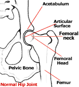
To understand hip dysplasia we must have a basic understanding of the joint that is being affected. The hip joint forms the attachment of the hind leg to the body and is a ball and socket joint. The ball portion is the head of the femur while the socket (acetabulum) is located on the pelvis. In a normal joint the ball rotates freely within the socket. To facilitate movement the bones are shaped to perfectly match each other; with the socket surrounding the ball. To strengthen the joint, the two bones are held together by a strong ligament. The ligament attaches the femoral head directly to the acetabulum. Also, the joint capsule, which is a very strong band of connective tissue, encircles the two bones adding further stability. The area where the bones actually touch each other is called the articular surface. It is perfectly smooth and cushioned with a layer of spongy cartilage. In addition, the joint contains a highly viscous fluid that lubricates the articular surfaces. In a dog with normal hips, all of these factors work together to cause the joint to function smoothly and with stability.
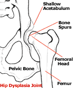
Hip dysplasia is associated with abnormal joint structure and a laxity of the muscles, connective tissue, and ligaments that would normally support the joint. As joint laxity develops, the articular surfaces of the two bones lose contact with each other. This separation of the two bones within the joint is called a subluxation, and this causes a drastic change in the size and shape of the articular surfaces. Most dysplastic dogs are born with normal hips but due to their genetic make-up (and possibly other factors) the soft tissues that surround the joint develop abnormally causing the subluxation. It is this subluxation and the remodeling of the hip that leads to the symptoms we associate with this disease. Hip dysplasia may or may not be bilateral; affecting both the right and/or left hip.

CHERRY EYE
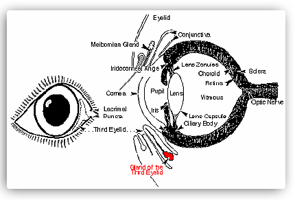
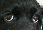
Cherry eye is the common name for a the condition caused when the gland of the third eyelid of the dog, otherwise known as Harder's gland or nictitans gland, becomes inflamed, swells up, pops out of place on the bulbar side of the third eyelid, and becomes more inflamed, swollen and irritated such that it becomes bloody and ulcerated, and can cover 1/2 to the whole eyeball of the dog. The condition is then referred to as follicular conjunctivitis. It used to be that when dogs developed cherry eye, veterinarians would simply remove the gland, just as medical doctors routinely removed inflamed tonsils in people. Within the past decade or so, it has now been decided that removing the gland is bad because perhaps the removal of that gland will result in reduced tear production leading to dry eye or keratoconjunctivits sicca. So now veterinarians routinely want to sew the prolapsed gland back in its original place on the third eyelid, sort of under the eyeball. There are other glands around the eyeball which make tears, and the gland of the third eyelid is not the major gland, but veterinarians are afraid of doing anything which might reduce tear production and cause the dry eye.
Read more: What Are the Causes of Cherry Eye? | eHow.com http://www.ehow.com/list_6077090_causes-cherry-eye_.html#ixzz1IkUzdAkt
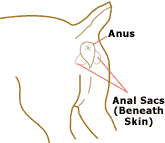
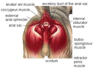
As the dog or cat is viewed from behind, anal glands (also called anal sacs) are located on each side of and slightly below the anal opening, at the 4 o'clock and 8 o'clock positions. A tiny duct or tube leads under the skin to an opening directly beside the anus.
Anal gland impactions, infections, and abscesses can occur. Here is how: For various reasons, such as the conformation of the animals, the thickness of the gland's secretions, or the softness of the stool, these glands and their ducts often become clogged, or 'impacted.' When this occurs, the animal will sit down on its rear quarters and drag its anal area across the floor or ground. This is called 'scooting.' Both dogs and cats may lick the anal area excessively. Impacted anal glands are a very, very common problem for dogs, especially the smaller breeds.
Anal glands may also become infected and abscess. Bacteria make their way into the glands, probably through the ducts. This is a very painful condition, and the first sign you may see is that the animal attempts to bite or scratch when you touch the area near the tail.
Treatment of anal gland problems is usually pretty straight forward. If the glands are impacted (backed up), they can be manually expressed (emptied). Your vet can do this, and he or she can show you how to do it so that you can do it yourself in the future if needed. Your vet will show you how to use your fingers or a thumb and forefinger to gently apply pressure on either side of the anal opening - on the outside of the glands. Press inwards and upwards and you should see the fluid being expressed.
Source: CanineJournal.com
Foods That are Poisonous for dog to eat